Cervical Spinal Cord Compression
Seven small vertebrae begin at the base of the skull and form the neck called the cervical spine. In between your vertebrae are flexible intervertebral disks. They act as shock absorbers when you walk or run.
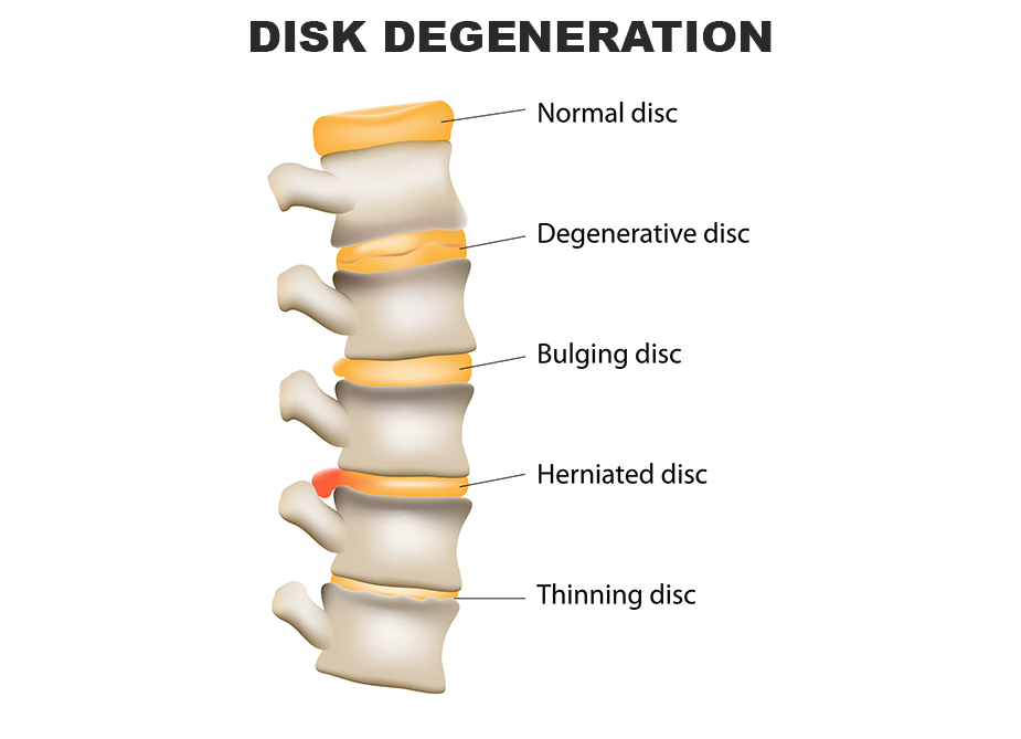
Intervertebral disks are flat and round and about a half inch thick. The disks are made up of a tough outer ring and a jelly-like center.
As the disks in the spine age, they lose height and water content. When they lose height, the vertebrae move closer together and become compressed. Cervical spinal cord compression causes a condition called cervical spondylotic myelopathy (CSM). Other causes of spinal cord compression are rheumatoid arthritis and neck injuries.
Nerves branch out from the spinal cord through openings in the vertebrae and carry messages between the brain and muscles all over your body.
Guests with CSM may experience a variety of symptoms such as:
- Weakness in the muscles of the arms, shoulders, or hands. Difficulty grasping.
- Loss of balance and coordination. Difficulty walking
- Loss of fine motor skills. Difficulty writing, buttoning your clothes, or handling a knife and fork.
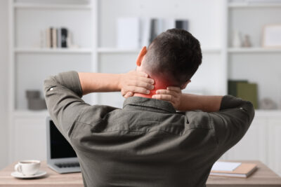
- Pain or stiffness in the neck
Several side affects of CSM are herniated or bulging disks and bone spurs:
- If the disk is degenerated and compressed, the jelly-like nucleus may squeeze all the way through the outer ring. When it bulges out toward the spinal canal, it can put pressure on the spinal cord and/or nerve roots.
- The body responds to a collapsed disk by forming bone spurs around the disk to add strength. The extra bone may narrow the spinal canal, compressing the spinal cord.
After an examination and discussion of symptoms, your DOC surgeon or PA will check for changes in your reflexes, numbness and weakness in arms hands, and legs, balance and coordination difficulties, and muscle deterioration. X-rays will show the alignment of the cervical vertebrae. An MRI shows spinal cord compression and damage to soft tissues, such as a bulging or herniated disk. A CT scan provides imaging details of the narrowing of the spinal canal and bone spurs. A myelogram is a special CT scan. Contrast dye is injected into the spinal column to enhance details of the spinal cord and nerve roots.
The goal of nonsurgical treatment is to decrease your pain and improve your ability to perform daily activities.
Nonsurgical treatment options include:
- Soft cervical collar to allow the muscles of the neck to rest and limit neck motion for a limited period of time.
- Physical therapy to help relieve pain, strengthen neck muscles and increase flexibility.
- Medications to relieve pain and inflammation.
If nonsurgical treatments fail to provide relief from pain and dysfunction, the DOC surgeon will discuss surgical options with you. In order to stop the progressive dysfunction of the spinal cord and restore your ability to enjoy everyday activities, cervical spine surgery that decompresses the spinal canal may be necessary.
For more information on the cost of care, click here